MCQ : Nuclear-25
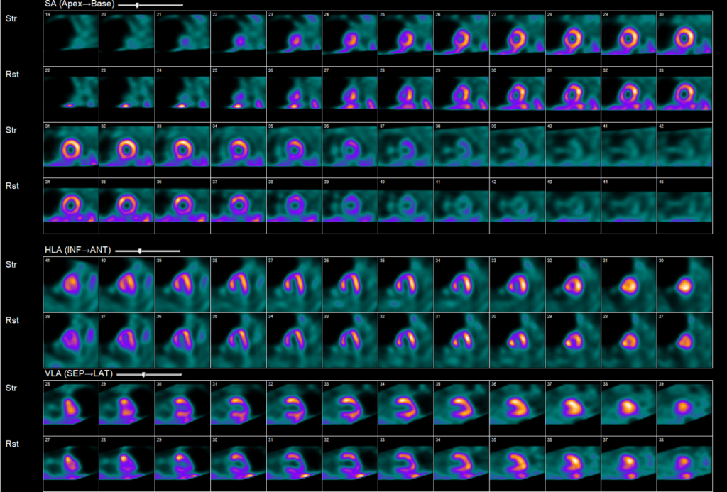
70 year old female with chest pain.
- Hypertension, Diabetes and poor mobility
- Rest ECG is normal
- Rests-stress regadenoson stress test was performed
What is the best interpretation of these images?
A) LAD territory infarct
B) LAD territory infarct and ischemia
C) Breast attenuation
D) Conduction abnormality
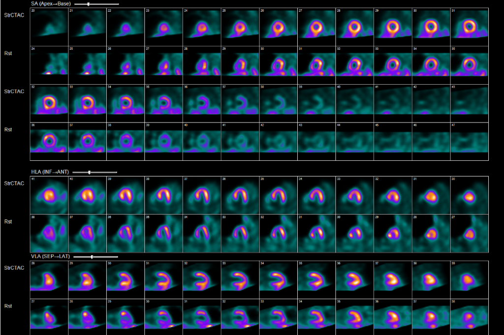
Stress images with CT attenuation correction
Correct Answer is C) : Breast attenuation
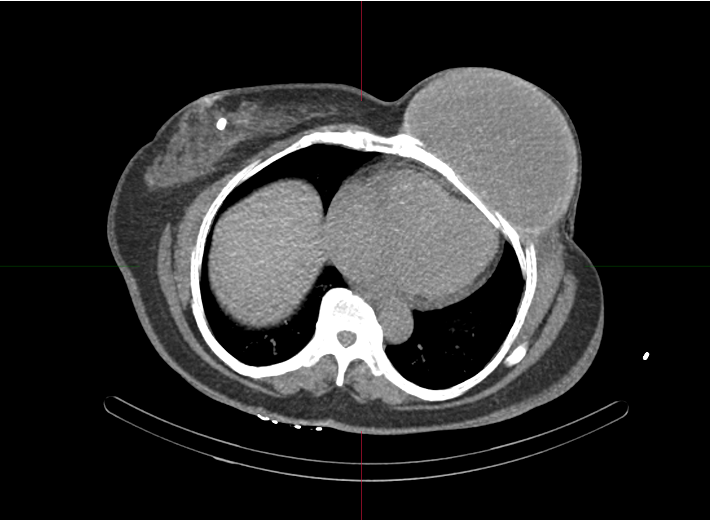
- •Prior mastectomy and breast reconstruction.
- •Note focal calcification in right breast.
Defects are not always ischemia/Infarct related.
- •Review RAW images
- •Use CT attenuation correction to overcome attenuation artifacts
- •Look at ECG to assess for LBBB or pacing artifacts.
![]()