MCQ : Nuclear-23
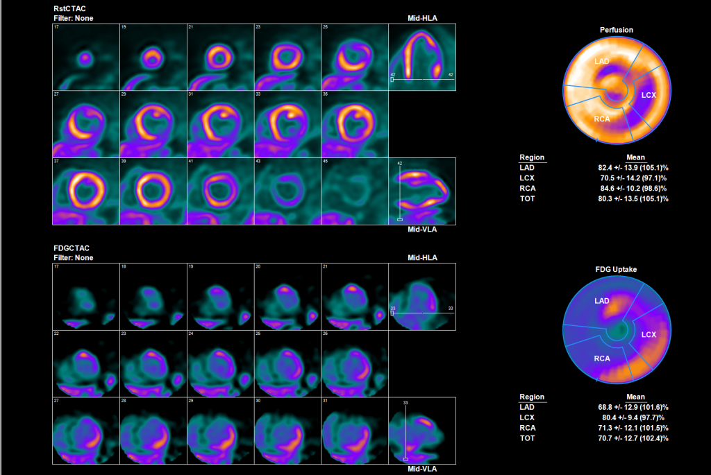
A 42 year old female who presents with polymorphic ventricular tachycardia. She was resuscitated and a coronary angiogram revealed evidence of mild coronary atherosclerosis. An echocardiogram showed normal valves and right ventricular systolic function with mildly reduced left ventricular ejection fraction at 41%.
A Rest Rb-82 perfusion with FDG-18 metabolic imaging was performed to rule out cardiac sarcoidosis.

The images are most consistent with:
A) Patchy scar but no inflammation
B) Patchy inflammation and no scar
C) Failure to suppress normal FDG uptake due to a poor diet preparation
D) LV cavity blood pool activity of FDG with no inflammation
Correct Answer is B) : Patchy inflammation and no scar
There is evidence of multiple perfusion defects (Top images) with enhanced FDG uptake in the same distribution as the perfusion defects (Bottom images). This perfect perfusion defects pattern with matched FDG uptake pattern is the whole-mark of identification of inflammation by PET.
Patient in addition had peri-hilar lymphnodes uptake which were biopsied and confirmed the presence of pulmonary sarcoidosis with now cardiac involvement.
![]()
