MCQ : CT-02
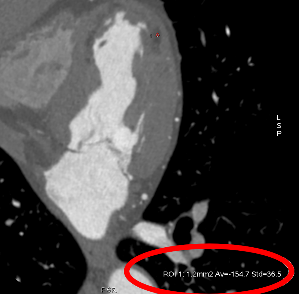
65 yo male, history of CAD with previous anterior myocardial infarction, undergoing CT for atypical chest pain.
What do you see?
A – Heavy calcifications on LAD and RCA, mixed plaque with moderate stenosis on LCX, left ventricular apical thrombus
B – Occluded LAD stent, patent RCA stent, mixed plaque with mild stenosis on LCX, myocardium of the left ventricular apex with calcifications
C – Patent left main, LAD and RCA stents, mixed plaque with moderate stenosis on LCX, left ventricular myocardial apex with calcifications
D – Patent left main, LAD and RCA stents, mixed plaque with moderate stenosis on LCX, myocardium of the left ventricle apex and anterior wall with signs of adipose metaplasia

Right answer is D.
Cardiac CT showed patent left main, LAD, and RCA stents, mixed plaque (calcific and fibrolipidic components) with moderate stenosis on LCX. The apex and the anterior wall of the left ventricle showed signs of the previous infarction with presence of fatty metaplasia.
Lipomatousmetaplasia of the myocardium is often seen following myocardial infarction. Myocardial fat appears in cardiac CT as focal areas of lower attenuation (with HU values below −20 HU) than normal myocardium.
![]()
