MCQ : CT-01
60 year old patient, undergoing CT for atypical chest pain.
What do you see at the left ventricular apex?
A – Left ventricular lipoma
B – Apical aneurism with partially calcified thrombus
C – Partially calcified Metastatic Tumor Mass
D – Calcified Myxoma
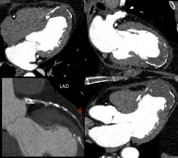
Right answer is B: Apical aneurism with partially calcified thrombus
Cardiac CT shows a left ventricle apical aneurysm with a huge, partially calcified thrombus (seen as a hypodense mass with lack of enhancement).
The aneurysm is the result of a previous anterior myocardial infarction due to occlusion of the left anterior descending artery (LAD), as illustrated in the panel.
![]()