MCQ : CMR-17
71-year-old male with history of no past past medical history
is presenting with dyspnea on exertion, lower extremities and abdominal fullness.

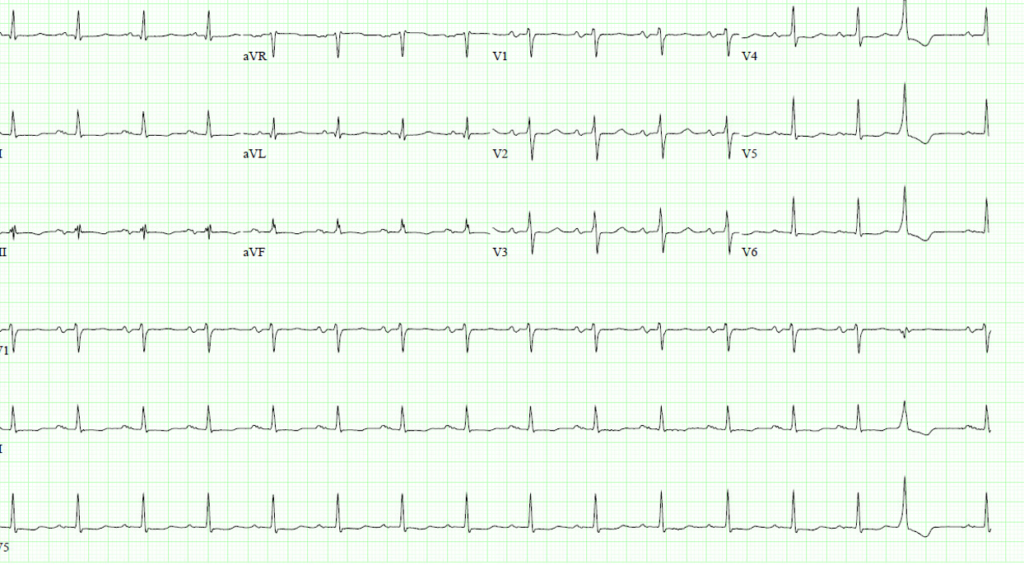
ECG: Sinus tachycardia and PVCs
MRI
Apical 4 chamber

IVC
Based on these images which is your diagnosis?
A) Severe LV dysfunction
B) Interventricular dependence
C) Severe RV dysfunction
D) Large pericardial effusion
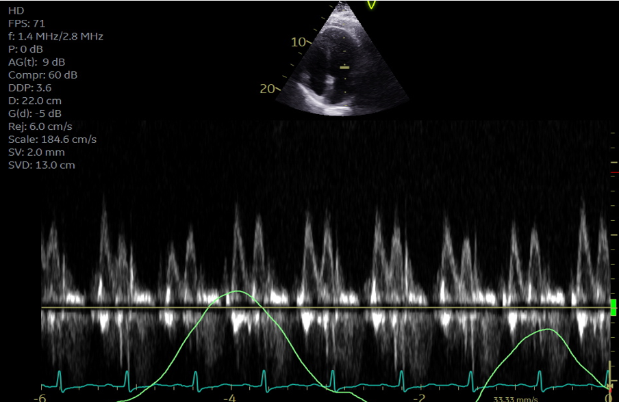
Correct answer is: B) Interventricular dependence with significant septal Bounce
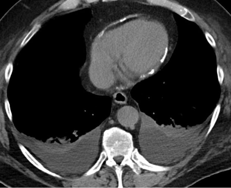
- Note significant pericardial thickening and calcifications on non contrast cardiac CT.
- Note presence pleural effusion.
- Diagnosis of pericardial constriction with calcified pericardium was made and patient was referred for pericardiectomy

- An abrupt anterior or posterior movement of the interventricular septum in early diastole (septal bounce or shudder) is commonly seen with constrictive pericarditis.
- Interventricular septal bounce with abrupt transient leftward movement of the interventricular septum with inspiration and rightward movement of the interventricular septum with expiration (resulting from ventricular interdependence). Note Doppler findings across MV supporting interventricular dependence.
- Note IVC plethora with no respiratory changes.
- This finding corresponds to the dip and plateau sign identified in invasive ventricular pressure recordings
- Increased pericardial thickness suggests constrictive pericarditis; presence or absence of pericardial thickening is not sufficient to establish or exclude the diagnosis
J Am Coll Cardiol Img. 2020 Jun, 13 (6) 1422–1437
![]()
