MCQ : Nuclear-07
67 year-old man, with previous MI
Recent onset of effort dyspnoea
ECG: inferior MI, VEB
Echo: poor acoustic window. EF: 35%
Waiting list for hip replacement
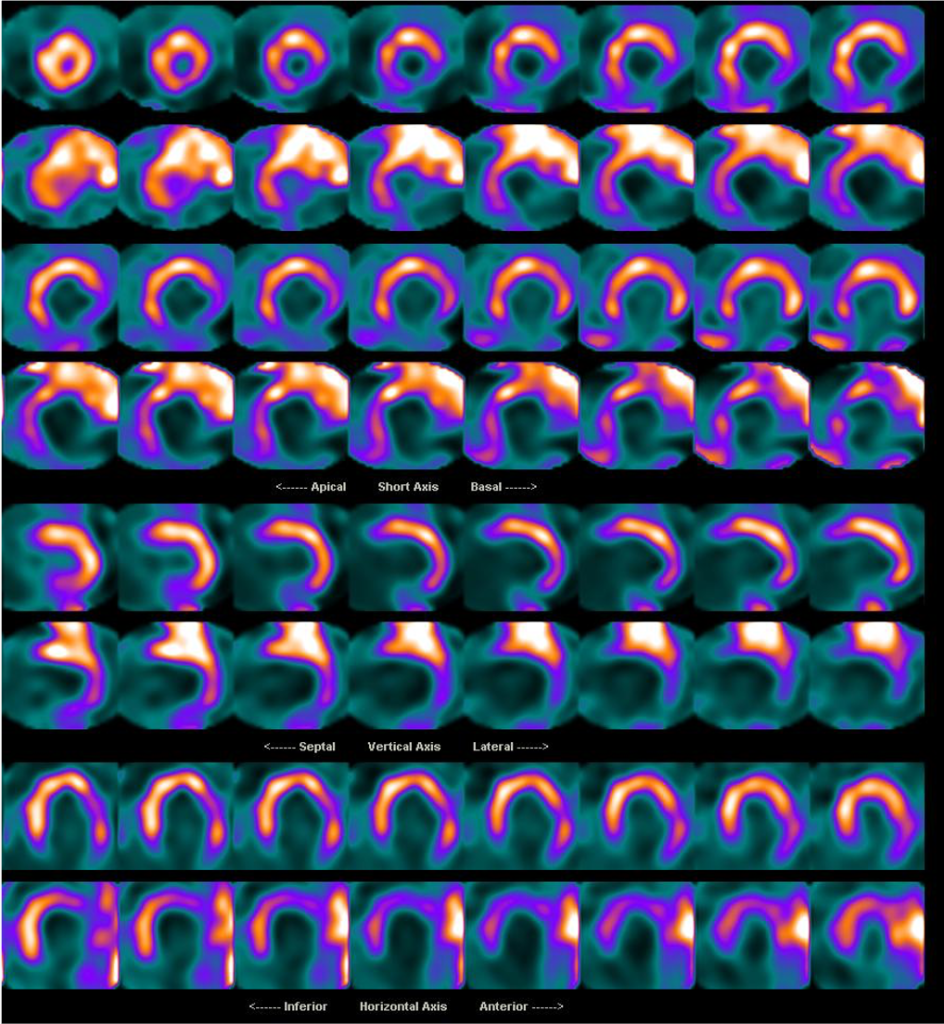
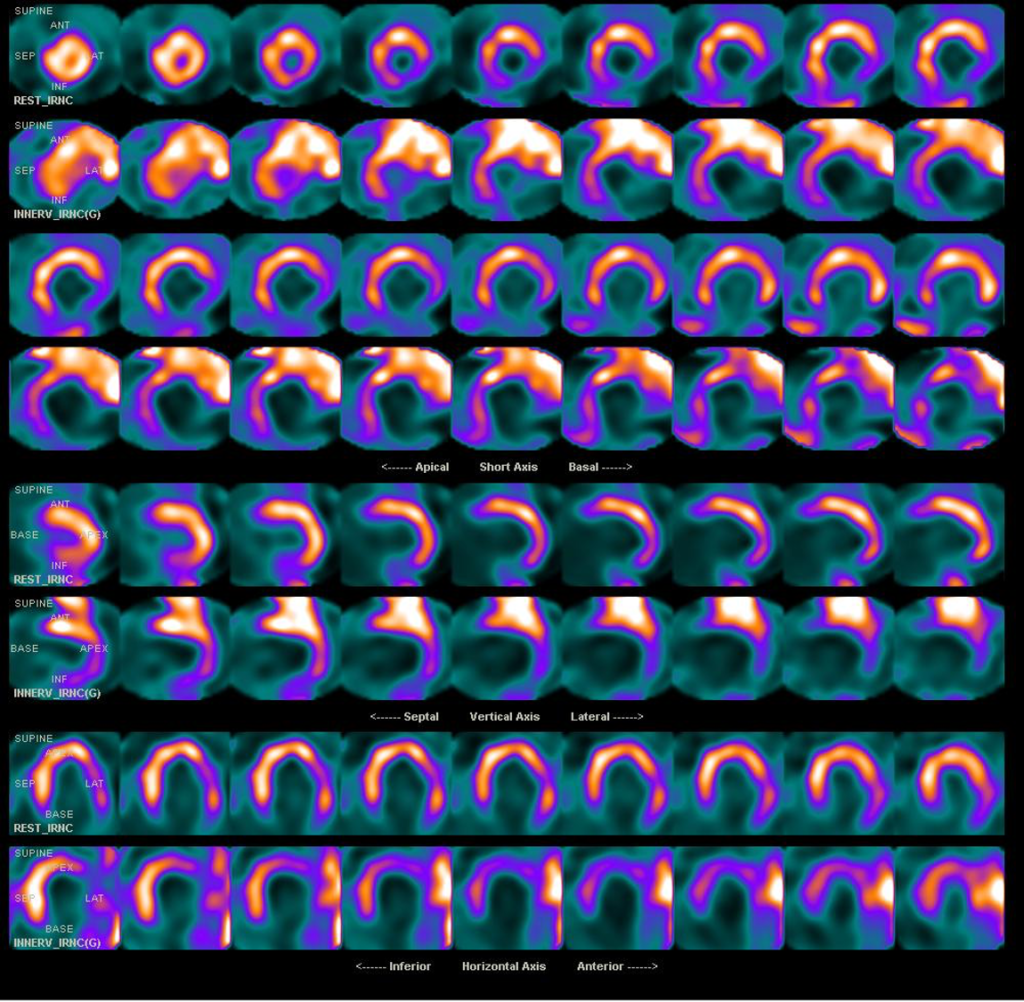
Which kind of tracer/s is/are used?
Which is the best interpretation of these images?
A) rest 201Thallium/ stress 99mTc Tetrofosmine, area of necrosis plus ischemia
B) 99mTc Tetrofosmine stress/rest study, area of necrosis plus ischemia
C) 99mTc Tetrofosmine/123I MIBG perfusion-innervation mismatch for the evaluation of EV area
D) 99mTc Tetrofosmine with and without nitrates study for the evaluation of viability


Right answer is: C) 99mTc Tetrofosmine/123I MIBG perfusion-innervation mismatch for the evaluation of EV area
Regional impairment of left ventricular (LV) sympathetic innervation, as evaluated at tomographic 123 I-MIBG imaging, has been demonstrated predicting adverse clinical events in patients with ventricular arrhythmias (VA)
In patients with VA submitted to EAM, successful ablation sites have been shown to co-localize at the level of perfusion/innervations mismatch, characterized by viable myocardial regions but impaired sympathetic innervations, which may represent possible targets during ablation
![]()
