Since April 2020, Cardiac Imaging Agorà is a free space, where experts in cardiac imaging share their knowledge and experience to improve patient’s care.
Tutorials
Visit our index pages to see the list of available videos. Tutorials are classified and provided both by modality and by disease.
Role of MMI for assessing CAD in women Alessia Gimelli, MD
Incidental findings on AC imaging: a radiologist’s perpective Michael Bolen, MD
Cardiac Imaging in Stable CAD: the broader perspective Richard Underwood, MD
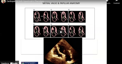
Cardiopathies with Hypertrophic Phenotype Georgette Khoury, MD
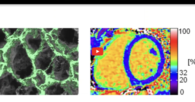
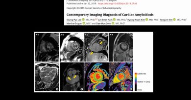
Imaging the Amyloidosis disease by CMR Marianna Fontana, MD
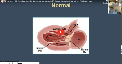
Hypertrophic Cardiomyopathy: Anatomic Variants and Echocardiographic Pearls from Dr Harry Lever Harry M. Lever, MD
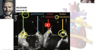
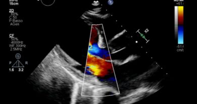
Echo To Guide MitraClip Procedure Özge Özden Tok, MD
The Results of the ISCHEMIA trial with Dr Leslee Shaw Leslee J. Shaw, MD
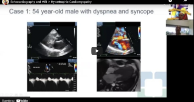
Echocardiography and MRI in Hypertrophic Cardiomyopathy Milind Y. Desai, MD
Cardiac CT tips and tricks Marco Guglielmo, MD
Echocardiography in identifying mechanical complications of myocardial infarction Venu Menon, MD
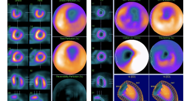
Step by Step reading a normal stress CZT MPS Alessia Gimelli, MD
PET vs. SPECT: Tracers , Equipment, protocols Wael Jaber, MD
Type of stress test in nuclear cardiology Alessia Gimelli, MD
Reading a Normal Cardiac PET Scan Wael Jaber, MD
MCQ of the Week
Since December 2020 Multiple Choice Questions are published weekly.see them all or see the following latest ones
79-year-old man subacute anterior myocardial infarction treated with PCI+DES on the mid left anterior descending artery.
What is the structure marked in this video?
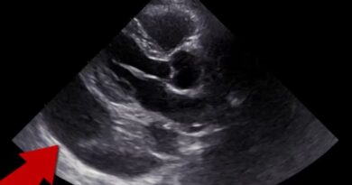
38 year old female with syncope. What is the best interpretation based on this image?
71-year-old male with history of no past past medical history is presenting with dyspnea on exertion, lower extremities and abdominal fullness.
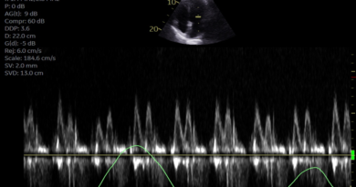
75-year-old male with history of HFrEF who walks in to your clinic for a follow-up Echocardiogram.
70 year old female with chest pain. What is the best interpretation of these images?
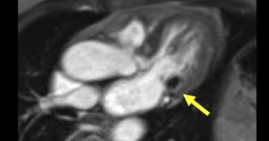
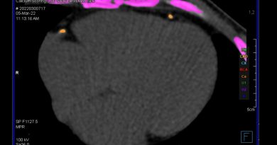
What do you think about the mass indicated by yellow arrow?
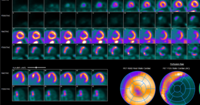
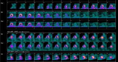
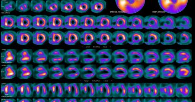
61 yo man. COVID-19 during April 2020. New onset of dyspnoea. BBSn. Echo evaluation showed EF 48% with hypokinesia of the apex. He was submitted to stress/rest MPS. What is the best interpretation of these images?
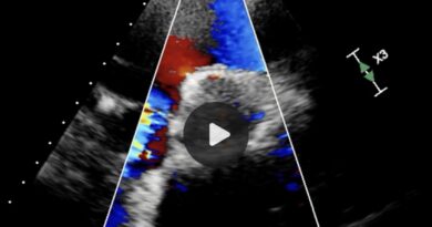
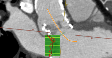
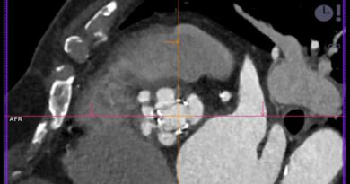
Pre Valve in mitral annulus calcification (MAC) assessment in a patient with severe mitral stenosis and previous TAVI
Haiku Imaging
Started on January 2022see them all or see the following latest ones
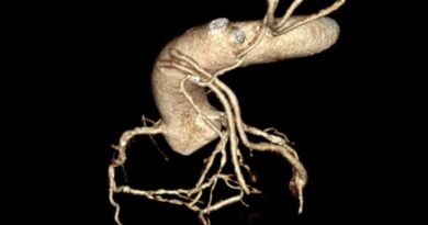
How to scan patients with CABG? Lucia La Mura, MD
Ischemic Mitral Regurgitation Ilaria Dentamaro, MD
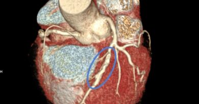
How to perform CT scan in patients with STENTs Lucia La Mura, MD
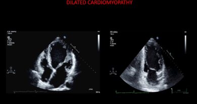
Echocardiography in dilated cardiomyopathy Ilaria Dentamaro, MD
The role of cardiac CT in the evaluation of prosthetic dysfunctions: a clinical case Lucia La Mura, MD
"Imaging" Cardiac Amyloidosis with CMR Ilaria Dentamaro, MD
How to perform a cardiac CT scan for TAVI Lucia La Mura, MD
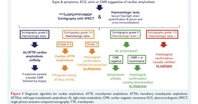
How to integrate bone scan in the clinical work out of cardiac amyloidosis Alessia Gimelli, MD
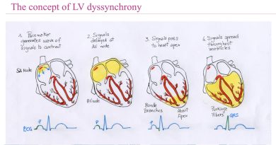
The role of nuclear cardiology in the evaluation of dyssynchrony Alessia Gimelli, MD
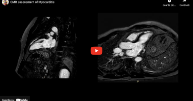
CMR assessment of Myocarditis Ilaria Dentamaro, MD
Cardiac Imaging Agorà is also on YouTube Channel at https://www.youtube.com/c/CardiacImagingAgora
![]()
![]()
